
VIP member
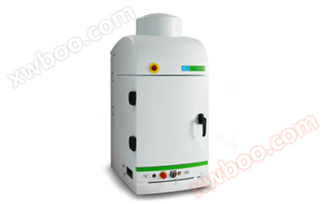
IVIS Lumina X5 Imaging System
PerkinElmer's next-generation small animal live imaging system IVIS Lumina X5 integrates high-throughput bioluminescence, fluorescence, and high-resol
Product details
- Product Description: PerkinElmer's next-generation small animal live imaging system IVIS Lumina X5 integrates high-throughput bioluminescence, fluorescence, and high-resolution 2D X-ray imaging modules. Expanded optical and X-ray imaging field of view to five mice, unique experimental setup, and sample labeling accessories
- Condensation temperature:
- Vacuum degree:
- Freeze dried area:
- Product model:
- Instrument type:
- Instrument material:
- Instrument structure:
- Heating power:
- Sample size:
- Test tube size:
- Heating method:
- Electrical resistivity:
- Pure water quality:
- Product type:
- Categories: Live Molecular Imaging, Life Science Laboratory Instruments
PerkinElmer's next-generation small animal live imaging system IVIS Lumina X5 integrates high-throughput bioluminescence, fluorescence, and high-resolution 2D X-ray imaging modules. Expanding the optical and X-ray imaging field to five mice, unique experimental settings, and sample labeling accessories allow researchers to more conveniently and quickly obtain stable data and answers on anatomical and functional aspects of disease progression.
IVIS Lumina X5 Imaging System
Part number: CLS148590
overview
In addition to all the functions of the IVIS Lumina S5 imaging system, the IVIS Lumina X5 also integrates high-resolution X-ray imaging capabilities. The ability to integrate high-resolution X-ray images with high-quality optical image data makes it an industry recognized two-dimensional multi-mode live imaging system. In addition, the IVIS Lumina X5 includes advanced spectroscopy and spectral separation capabilities, which can monitor multiple biological events within the same animal through high-sensitivity multispectral imaging.
Thoroughly high-throughput optics andX-ray live imaging system
The IVIS Lumina X5 integrates a one inch CCD camera on the classic Lumina platform, providing a high-throughput imaging field of 20 x 20 centimeters, sufficient for simultaneous bioluminescence and fluorescence imaging of five mice. In addition, the new scintillation screen can provide corresponding X-ray structural image data for any optical image within the imaging field. In addition, the automatic imaging and independently adjustable height scintillation screen design allows researchers to conveniently perform accurate optical/X-ray fusion imaging on 500-600 gram rats.
Like other IVIS Lumina imaging systems, the X5 system is equipped with 26 filters suitable for imaging fluorescence signals ranging from green to near-infrared. The new excitation technology effectively extends the fluorescence imaging range to 900 nm. In addition, the IVIS Lumina X5 system integrates PerkinElmer's Computational Pure Spectroscopy (CPS) algorithm and spectral database generation software tool, enabling accurate removal of spontaneous fluorescence background, multi signal splitting, and fluorescence signal quantification functions.
The quantitative function of the IVIS system has been recognized by the industry. The system's calibration function enables consistent and reproducible data results to be obtained under different magnification factors, filter selections, or camera parameter settings. In addition, whether the data comes from the same instrument or instruments from other research institutions, as long as they are all on the IVIS platform, the data can be directly compared with each other.
Industry leadingX-ray imaging resolution
The IVIS Lumina X5 is equipped with a microfocus X-ray light source, which greatly improves the resolution of X-ray imaging. With the help of high X-ray imaging resolution, researchers can not only clearly understand the anatomical changes of organs during the development of diseases, but also accurately locate the optical observation objects, thus more comprehensively interpreting diseases.
Rich selection of accessories- Simplify the experimental operation process
The IVIS Lumina X5 not only expands imaging flux by upgrading the large-sized CCD camera, but also comes equipped with various imaging accessories (separately optional) for pre operation of experimental animals before imaging, helping users conduct experiments more conveniently.
Before imaging begins, users can place the experimental animals in the experimental animal tray in advance and load them into the imaging darkbox as a whole, without the need to perform the above operations in the experimental darkbox as before. At the same time, the imaging software can automatically identify and record the experimental animals at the corresponding positions based on the reference labels built into the tray, and perform ROI circle selection quantification.
The new animal tray adopts a modular design, and each module can be easily disassembled for easy cleaning. The animal anesthesia interface in the tray also adopts a nose less cone design for easy cleaning. In addition, combined with the powerful vacuum circulation device of the new generation gas anesthesia machine RAS-4, the degree of anesthesia gas leakage is minimized, improving the safety of use.
New Living Image ® The imaging and analysis software also has built-in animal ID recognition function. By combining the use of third-party animal ID chip recognition technology, users can easily identify, record, and output images and quantitative data from different experimental animal individuals.
Main features:
- High throughput optical and X-ray imaging (simultaneously imaging 5 mice)
- High resolution, low radiation X-ray imaging
- Support imaging of mice and rats
- High sensitivity bioluminescence imaging
- High sensitivity bioluminescence imaging
- High sensitivity bioluminescence imaging
- Bioluminescence, fluorescence, and X-ray multimodal imaging
- Rich imaging and data analysis accessories
specifications
|
Imaging mode |
Optical Imaging |
|
Product brand name |
IVIS |
attribute
Online inquiry
-
Contacts
-
Company
-
Telephone
-
Email
-
WeChat
-
Verification Code
-
Message Content
-