
VIP member
FluorCam Multispectral Fluorescence Imaging System
FluorCam Multispectral Fluorescence Imaging System
Product details
FluorCam Multispectral Fluorescence Imaging System
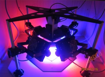
 FluorCamThe multispectral fluorescence imaging system is an advanced extension product of FluorCam chlorophyll fluorescence imaging technology, which can be used for both chlorophyll fluorescence dynamic imaging analysis and other applicationsLong wavelength UV light (320nm -400nm)Triggered by plant leavesMultispectral fluorescence imaging measurement and analysis, and imaging measurement of steady-state fluorescence such as green fluorescent protein GFP can also be optionally selected. The maximum imaging area of the standard configuration (standard version) is13 x 13 cmThe maximum imaging area of the large version is20 x 20 cmWidely used in plant photosynthetic physiology and ecology, plant stress physiology and susceptibility, stomatal function, plant environment such as soil heavy metal pollution response and biological detection, plant resistance, crop breeding Phenotyping、 Research on genetic modification, steady-state fluorescence imaging measurement, etc.
Functional features:
üMulti excitation luminescence multispectral fluorescence imaging technologyThrough optical filter technology, only specific wavelengths of light (excitation light) are allowed to reach the sample to excite fluorescence, while only specific wavelengths of excitation fluorescence are allowed to reach the detector. Different fluorescent chromophores (such as chlorophyll or GFP green fluorescent protein) are "sensitive" to excitation light of different wavelengths and absorb it to excite fluorescence of different wavelengths. Based on this principle, two or more excitation light sources, green wheels, and corresponding filters can be selected to image and analyze fluorescence of different wavelengths (multispectral fluorescence). If red light, blue light, and corresponding filters are selected, GFP and chlorophyll fluorescence imaging analysis can be performed. Green light source and corresponding filters can also be selected for fluorescence imaging analysis of YFP;
ü

UV induced multispectral fluorescence imaging technologyLong wavelength UV light (320nm -)400nm)Excitation of plant leaves can produce a fluorescence spectrum with four characteristic peaks, namely blue light 440nm (F440), green light 520nm (F520), red light 690nm (F690), and far-infrared 740nm (F740). F440 and F520 are collectively referred to as BGF, which is emitted from the epidermis, mesophyll cell walls, and leaf veins. F690 and F740 are chlorophyll fluorescence Chl-FUV induced multispectral fluorescence can be used to sensitively and specifically evaluate plant physiological states, including stress states such as drought, pests and diseases, environmental pollution, nitrogen stress, etc
üThe imaging area is large, with a standard configuration of 13x13cm. The large version has an imaging area of 20x20cm, which can be used for experimental imaging analysis of the entire plant or even multiple plants (such as small plants like Arabidopsis)
üIt can perform automatic repeated imaging measurement and unmanned monitoring, and can set up two experimental programs (Protocols) to automatically cycle imaging measurement. The imaging measurement data is automatically stored in the computer according to the time date (with timestamp)
üEquipped with various universal experimental protocols such as Kautsky induction effect, fluorescence quenching analysis, GFP steady-state fluorescence imaging, and UV excited multispectral fluorescence imaging analysis, measuring and analyzing over 60 parameters
üOptional TetraCam color imaging module with a maximum imaging area of 20x25cm, used for morphological measurement and analysis. Combined with fluorescence imaging analysis parameters, it can be used as a powerful tool for plant phenotype analysis
üMeasurement samples include leaves, flowers, fruits, roots, other plant tissues, whole plants, algae, small animals, etc
Configuration specifications:.png")
1)Standard configuration: capable of chlorophyll fluorescence imaging analysis and fluorescence imaging analysis of 4 bands excited by UV light source, with an imaging area of 13 x 13cm. The system is highly integrated and convenient to use, equipped with a 7-bit green wave wheel and multispectral fluorescence imaging filter group, high-resolution CCD lens, UV UV excitation multispectral fluorescence imaging function module and program software, etc;
2)Expansion configuration: Modular structure with high scalability, not only capable of chlorophyll fluorescence imaging analysis and fluorescence imaging analysis of four bands excited by UV light source, but also capable of PAR absorption and NDVI imaging analysis, GFP green fluorescent protein and other steady-state fluorescence imaging analysis (optional). Different excitation light source and filter combinations can be selected according to customer needs, with an imaging area of 13 x 13cm;
3)Large scale configuration: It has all the functional advantages of the above extended configuration, with an imaging area of 20cm x 20cm, and can perform imaging analysis on the entire plant or multiple plants.
Technical indicators:
ØThe standard version has an imaging area of 13x13cm, while the large version has an imaging area of 20x20cm
ØThe standard configuration includes a filter wheel, ChlF filter, and high-resolution lens, with measurement parameters including Fo, Fo’, Fs, Fm, Fm’, Fp, FtDn, FtLn, Fv, NPQ_Dn, NPQ_Ln, Qp_Dn, Qp_Ln, qN, QY,QY_Ln, PARabs, RMore than 60 chlorophyll fluorescence parameters and steady-state fluorescence, including fd, BGF, UV-Chl. F, etc
ØUV excited multispectral fluorescence parameters include F440, F520, F690, F740
ØHigh resolution TOMI-2 CCD sensor
a)Line by line scanning CCD
b)Maximum image resolution: 1360 × 1024 pixels
c)Time resolution: up to 20 frames per second at the highest image resolution
d)A/D conversion resolution: 16 bits (65536 grayscale levels)
e)Pixel size: 6.45 µ m × 6.45 µ m
f)Operation mode: 1) Dynamic video mode, used for measuring chlorophyll fluorescence parameters; 2) Snapshot mode, used for measuring fluorescent proteins such as GFP and fluorescent dyes
g)Communication mode: Gigabit Ethernet
ØAutomatic measurement and analysis function (unmanned): can be preset1A or2A test program, the system can automatically measure and store, such as automatic timed operation during the dayKautskyInduction effect program, automatic timed running of fluorescence quenching analysis program at night
ØEquipped with universal chlorophyll fluorescence imaging measurement protocols and steady-state fluorescence measurement protocols (optional), including Fv/Fm Protocol, Kautsky induction effect protocol, fluorescence quenching analysis protocol, steady-state fluorescence measurement, customer customized light response curve and PAR absorption imaging measurement, etc
Ø4individual13x13cmLEDLight source board (large version is 20x20cm), dual color light source(2Red Orange Light+2Blue light, dual color chemical light(Actinic light1andActinic light2)
ØStandard configurationActinic1light intensity300µmol(photons)/m².s,Actinic2light intensity2000µmol(photons)/m².sSaturated flicker4000µmol(photons)/m².s
ØLight source upgrade:Actinic1light intensity2000µmol(photons)/m².s,Actinic2light intensity3000µmol(photons)/m².sSaturated flicker6000µmol(photons)/m².s
ØThe standard measurement light is 618nm red light, with other bands available for selection and durationAdjustable from 10 µ s to 100 µ s;
Ø7-bit filtering wheel and filter, used for imaging measurement of chlorophyll fluorescence, F440, F520, F690, F740, and steady-state fluorescence such as GFP (GFP fluorescence requires corresponding functional modules to be selected)
ØOptional 735nm (FAR) and 630nm dual color LED light source boards with corresponding filters and functional program modules for measuring Fo’、 PARabs and NDVI
ØOptional 1 pair of blue LED light source board and corresponding filter, light intensity 3000µmol(photons)/m².sUsed for the measurement and research of stomatal function
ØOptional 1 pair of green LED light source boards for measuring YFG (corresponding filters must be selected)
ØIf measuring other fluorescence parameters, corresponding filters must be selected (please consult EcoLab laboratory). The following are selection references:

ØFluorCam chlorophyll fluorescence imaging analysis software, with Live (live testing), Protocol (experimental program selection) Pre-processing(Imaging Preprocessing), Result (Imaging Analysis Results) and other menus
ØThe Protocol experimental program can be freely edited, and customers can also use the wizard program template in the Protocol menu to freely create new experimental programs
ØImaging preprocessing can automatically select or manually select regions of interest (ROI) of different shapes, quantities, and positions. Imaging analysis results include high-resolution fluorescence dynamic images, histograms, imaging images with different parameters, and lists of fluorescence parameters for different ROIs
ØData analysis has "signal calculation and re averaging" mode (arithmetic mean) and "signal average and re calculation mode". In high signal-to-noise ratio situations, the "signal calculation and re averaging" mode is selected, and in low signal-to-noise ratio situations, the "signal average and re calculation" mode is selected to filter out errors caused by noise
ØGive Light System:Static or dynamic (sinus style)
ØBios:Firmware upgradable
ØCommunication method:Gigabit Ethernet
ØSupply voltage:90 – 240 V

Place of Origin:Europe
Online inquiry
-
Contacts
-
Company
-
Telephone
-
Email
-
WeChat
-
Verification Code
-
Message Content
-